
Архів офтальмології та щелепно-лицевої хірургії України Том 1, №2, 2024
Вернуться к номеру
Доцільність проведення кератотопографії у пацієнтів з тонкою рогівкою при підозрі на глаукому (клінічний випадок)
Авторы: Жмурик Д.В. (1, 2), Новак Л.П. (1, 2), Васильцов І.А. (1, 2), Шевчук Л.О. (1, 2)
(1) - Національний медичний університет імені О.О. Богомольця, м. Київ, Україна
(2) - Офтальмологічний медичний центр «OCHI Clinic», м. Київ, Україна
Рубрики: Хирургия, Офтальмология
Разделы: Справочник специалиста
Версия для печати
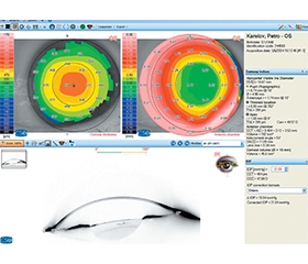
Цільовий внутрішньоочний тиск (ЦВОТ) — це індивідуально визначений рівень внутрішньоочного тиску (ВОТ), якого прагнуть досягти у пацієнтів з глаукомою або іншими захворюваннями, що супроводжуються підвищеним ВОТ, для запобігання прогресуючому пошкодженню зорового нерва і збереження зору. Визначення ЦВОТ базується на кількох факторах, включно з початковим рівнем ВОТ, який напряму корелює з даними пахіметрії. Пахіметрію можна проводити за допомогою різних апаратів, як-от The Argos Biometer Alcon, кератотопограф Schwind Sirius+ та пневмотонометр NCT-200. На прикладі наведеного клінічного випадку автори дійшли висновку, що найбільш вірогідні результати пахіметрії у пацієнтів зі зміненою рогівкою були отримані при використанні кератотопографа Schwind Sirius+. Завдяки отриманим даним була сформована відповідна тактика лікування, що дозволила стабілізувати ВОТ та прогресування наявних глаукомних змін.
Target intraocular pressure (IOP) is an individually determined level of IOP aimed to be achieved in patients with glaucoma or other diseases accompanied by high IOP to prevent progressive damage to the optic nerve and preserve vision. The determination of IOP is based on several factors, including baseline IOP, which directly correlates with pachymetry data. Pachymetry can be done using various devices such as Argos Biometer (Alcon), Schwind Sirius+ keratotopograph, and NCT-200 pneumotonometer. Based on this clinical case as an example, the authors concluded that the most reliable pachymetry results in patients with altered corneas were obtained on Schwind Sirius+ keratotopograph. Thanks to the findings, the appropriate treatment strategy was developed, which helped stabilize IOP and the progression of existing glaucomatous changes.
внутрішньоочний тиск; пахіметрія; кератотопографія; цільовий тиск
intraocular pressure; pachymetry; keratotopography; target pressure
Для ознакомления с полным содержанием статьи необходимо оформить подписку на журнал.
- Sedaghat MR, Moghaddam HM, Yekta AA, et al. Biomechani–cally-Corrected Intraocular Pressure Compared to Pressure Measured with Commonly Used Tonometers іn Normal Subjects. Clini–cal Optometry. 2019;11:127-133. http://doi.org/10.2147/OPTO.S220776.
- Silva FD, Lira M. Intraocular pressure measurement: A review. Survey of Ophthalmology. 2022;67(5);1319-1331. DOI: 10.1016/j.survophthal. 2022.03.001.
- Nuyen B, Mansouri K. Fundamentals and Advances in Tono–metry. Asia Pac J Ophthalmol (Phila). 2015 Mar-Apr;4(2):66-75. doi: 1097/APO.0000000000000118.
- Salvetat ML, Zeppieri M, Tosoni C, Brusini P. Repeatability and accuracy of applanation resonance tonometry in healthy subjects and patients with glaucoma. Acta Ophthalmol. 2014;92(1):66-73. doi: 1111/aos. 12209.
- Bao F, Deng M, Wang Q, et al. Evaluation of the relationship of corneal biomechanical metrics with physical intraocular pressure and central corneal thickness in ex vivo rabbit eye globes. Exp Eye Res. 2015;137:11-17. doi: 10.1016/j.exer.2015.05.018.
- Pronin S, Brown L, Megaw R, Tatham AJ. Measurement of intraocular pressure by patients with glaucoma. JAMA Ophthalmol. 2017;135(10):1030. doi: 10.1001/jamaophthalmol.2017.3151.
- Brown L, Foulsham W, Pronin S, Tatham AJ. The influence of corneal biomechanical properties on intraocular pressure measurements using a rebound self-tonometer. J Glaucoma. 2018;27:511-518. doi: 10.1097/1JG.0000000000000948.
- Martínez-Abad A., Piñero D.P. Pellucid marginal degene–ration: Detection, discrimination from other corneal ectatic disorders and progression. Cont Lens Anterior Eye. 2019;42(4):341-349. DOI: 10.1016/j.clae.2018.11.010.
- Mounir A. An Easy Guide to Pentacam Corneal Tomographgy. 2020. Available at: https://www.innovationinfobooks.com/ebookde–tails/an-easy-guide-to-pentacam-corneal-tomography [Accessed 11 August 2020].
- Motlagh MN, Moshirfar M, Murri MS, et al. Pentacam® corneal tomography for screening of refractive surgery candidates: A review of the literature, part I. Med Hypothesis Discov Innov Ophthalmol. 2019;8(3):177-203.