
Архів офтальмології та щелепно-лицевої хірургії України Том 1, №2, 2024
Вернуться к номеру
Можливості щодо деформації м’яких тканин різних ділянок голови при одновісному розтягненні
Авторы: D.S. Avetikov, K.P. Lokes, O.S. Ivanytska
Poltava State Medical University, Poltava, Ukraine
Рубрики: Хирургия, Офтальмология
Разделы: Клинические исследования
Версия для печати
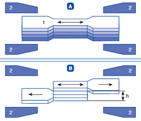
Актуальність. При бурхливому розвитку пластичної та реконструктивної хірургії наукові дослідження щодо вдосконалення оперативних методик в Україні практично не проводяться, оскільки це передбачає експерименти, створення контрольних груп і їхній комплексний аналіз. Усі сучасні методики пластичних реконструктивно-відновлювальних та естетичних операцій спрямовані на мобілізацію, тобто на деформацію (розтягнення й релаксацію) клаптів різної товщини з відшаруванням їх від підлеглих тканин. При цьому практично не проводилося досліджень щодо обґрунтування методик пластичних операцій, при яких окремі шари м’яких тканин голови відшаровуються на обмеженій ділянці або залишаються інтактними. Мета: визначити ступінь деформації м’яких тканин у різних ділянках голови залежно від її форми, віку пацієнта, статі й часу деформації і встановити зони з однаковими характеристиками деформації. Матеріали та методи. На першому етапі біомеханічних досліджень використовували розривну машину ZM-20. Один кінець фіксували в нерухомій частині затискача, другій — у рухомій. Результати. Найменшу пластичну деформацію мають м’які тканини носової ділянки, що слід враховувати на етапі планування операцій при усуненні патологічних рубців і рубцевих деформацій у цій зоні. Найбільшу пластичну деформацію мають привушно-жувальна ділянка, нижній край виличної, що прилягає до привушно-жувальної, та медіальний край очноямкової зони. Для цих ділянок характерне депонування жирової клітковини між дермою і поверхневою фасцією. Остання при цьому втрачає багатошарову будову та є пластинкою сполучної тканини, що оточує пучки мімічних м’язових волокон. Висновки. Виявлено суттєві відмінності у ступені деформації м’яких тканин голови залежно від антропометричних даних, віку, статі та часу деформації. Найбільш варіабельними є абсолютні значення пластичної деформації тканин щічної ділянки, що залежать від віку та статі: у чоловіків 41–50 років Ε = 0,54 (0,79–0,32) при m = 0,068, у жінок 51–65 років Ε = 0,54 (0,68–0,31) при m = 0,069. Найменша залежність від форми голови зафіксована в чоловіків-доліхоцефалів: Ε = 0,61 (0,79–0,38) при m = 0,077. Найменшу пластичну деформацію мають м’які тканини носової ділянки в брахіцефалів: Ε = 0,53 (0,72–0,32) при m = 0,062.
Background. With the rapid development of plastic and reconstructive surgery, scientific research on the improvement of surgical methods is practically not carried out in Ukraine, since they mean experiments, the creation of control groups and their comprehensive analysis. All modern methods of plastic reconstructive and aesthetic surgery are aimed at mobilization, i.e. at deformation (stretching and relaxation) of flaps of different thickness with their detachment from underlying tissues. At the same time, there were practically no studies on the substantiation of plastic surgery techniques, in which individual layers of soft tissues of the head are peeled off in a limited area or kept intact. Purpose: to determine the degree of deformation of soft tissues in different areas of the head depending on its shape, patient’s age, gender and time of deformation and to establish areas with the same characteristics regarding their deformation. Materials and methods. We used a ZM-20 breaking machine to conduct biomechanical research at the first stage. One end was fixed in a fixed grip, the other — in a moving grip. Results. The soft tissues of the nasal area are characterized by the smallest plastic deformation. This should be remembered at the stage of surgical planning when eliminating pathological scars and cicatricial deformations in this area. The greater plastic deformation is observed in the parotid-masticatory area, the lower margin of the zygomatic area adjacent to the parotid-masticatory area and the medial margin of the orbital area. These regions are characterized by deposition of fatty tissue between the dermis and superficial fascia. At the same time, the latter loses its multi-layered structure and is a plate of connective tissue surrounding bundles of mimic muscle fibers. Conclusions. Significant differences in the degree of deformation of the soft tissues of the head were found depending on anthropometric data, age, gender, and time of deformation. The most variable are the absolute values of plastic deformation of the tissues of the buccal area, which depend on age and gender: in men 41–50 years old, Ε = 0.54 (0.79–0.32), with m = 0.068, in women 51–65 years old, Ε = 0.54 (0.68–0.31), with m = 0.069. The smallest dependence was recorded on the shape of the head in dolichocephalic men: Ε = 0.61 (0.79–0.38), with m = 0.077. The soft tissues of the nasal area in brachycephals are characterized by the smallest plastic deformation: Ε = 0.53 (0.72–0.32), with m = 0.062.
пластична хірургія; щелепно-лицева ділянка; біомеханіка; м’які тканини; шкірно-жирові клапті
plastic surgery; maxillofacial area; biomechanics; soft tissues; skin-fat flaps
Для ознакомления с полным содержанием статьи необходимо оформить подписку на журнал.
- Uchida T., Kin T., Saito T., Shono N., Kiyofuji S., et al. De-Identification Technique with Facial Deformation in Head CT Images. Neuroinformatics. 2023. 21(3). 575-587.
- Langsdon P.R., Schroeder R.J. 2nd. Recognizing, Managing, and Guiding the Patient Through Complications in Facial Plastic Surgery. Facial Plast Surg Clin North Am. 2020. 28(4). 483-491.
- Nicksic P.J., Farmer R.L., Poore S.O., Rao V.K., Afifi A.M. Dermatologic Complications Following Cosmetic and Reconstructive Plastic Surgery: A Systematic Review of the Literature. Aesthetic Plast Surg. 2021. 45(6). 3005-3018.
- Lokes K.P., Voloshyna L.I., Ivanytska O.S., Yatsenko I.V., Gavriliev V.M., Rozkolupa O.O., Avetikov D.S. Formation of optimal scar on the background of surgical treatment of maxillofacial diseases of different origins. World of Medicine and Biology. 2023. 19(84). 102-105.
- Adidharma W., Chung K.C. Recent Advances in Upper Extremi–ty Microsurgery: From Traditional to Perforator Flaps. Hand Clin. 2024. 40(2). 161-166.
- Riopelle A.M., Jeong D., Boyd A.Y., Schanbacher C.F. Reconstruction of High-Tension Scalp Defects by the Twizzler Technique: A Retrospective Case Series. Dermatol Surg. 2023. 49(9). 832-837.
- Cameron M.B. Zachary, Grushchak S., Newman J. Skin Ana–tomy and Analysis. Facial Plast Surg Clin North Am. 2023. 31(4). 433-442.
- Basta M.N., Rao V., Paiva M., Liu P.Y., Woo A.S., Fi–scher J.P., Breuing K.H. Evaluating the Inaccuracy of the National Surgical Quality Improvement Project Surgical Risk Calculator in Plastic Surgery: A Meta-analysis of Short-Term Predicted Complications. Ann Plast Surg. 2022. 88(3). 219-223.
- Chen B., Genovese K., Pan B. In vivo panoramic human skin shape and deformation measurement using mirror-assisted multi-view digital image correlation. J Mech Behav Biomed Mater. 2020. 110. 103936.
- Yoo S., Kim M.R., Kim T.Y., Hwang S.J., Lim J.M., Park S.G. Relationship of transcutaneous oxygen tension with age and skin elasticity in Korean women. Skin Res Technol. 2020. 26(3). 325-328.
- Cotofana S., Fratila A.A., Schenck T.L., Redka-Swoboda W., Zilinsky I., Pavicic T. The Anatomy of the Aging Face: A Review. Facial Plast Surg. 2016. 32(3). 253-260.
- Arne Gerber P., Filler T. Static and Dynamic Anatomy of the Face, in Particular Eyebrows, Eyelids and Lips. Curr Probl Dermatol. 2022. 56. 306-312.
- Hu H., Shi Y., Qian Y., Yu X., Liu A., Li F., Jiang H., Wang H. Pyramidal multiple-theory (multi-type, multi-method and multi-layer) for facial fat grafting. J Cosmet Dermatol. 2023. 22(3). 937-944.
- Cotofana S., Lachman N. Anatomy of the Facial Fat Compartments and their Relevance in Aesthetic Surgery. J Dtsch Dermatol Ges. 2019. 17(4). 399-413.
- Wan M., Zhang J.X., Ding Y., Jin Y., Bedford J., et al. High-Risk Plastic Surgery: An Analysis of 108,303 Cases From the American College of Surgeons National Surgical Quality Improvement Program (ACS NSQIP). Plast Surg (Oakv). 2020. 28(1). 57-66.